

/ via embl/
Scientists used 3D images of the human nucleolus to visualise ribosome assembly pathways inside cells for the first time
Summary
- EMBL researchers have visualised the human nucleolus, the ‘factory’ for ribosome production, by combining cryo-electron tomography (cryo-ET) with correlative light and electron microscopy (CLEM).
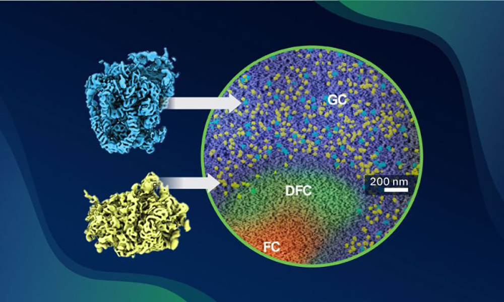
- The in-cell snapshots provided secondary structural detail for the two key ribosomal subunit precursors, the SSU processome and the pre-60S, and revealed new assembly configurations.
- The team found that a protein factor long thought to bind late in assembly actually binds much earlier, and that ribosomes do not mature in a linear assembly line from the centre of the nucleolus to its edge.
- The approach offers new ways to study large molecular complexes in the crowded environment of the nucleus.
For decades, ribosomes have been, quite literally, under the lens of many scientists. Ribosomes are highly sophisticated and complex molecular machines that allow cells to synthesise proteins. However, putting ribosomes together requires intricate assembly processes. While many underlying mechanisms for building ribosomes have been discovered ex situ (outside their native cellular context), our understanding of how ribosomes are assembled in situ has so far remained limited.
Through a collaboration between the research groups of Christoph Müller, Sara Cuylen-Häring and Julia Mahamid at EMBL Heidelberg, researchers have now pushed these limits. By combining cryo-electron tomography (cryo-ET) with correlative light and electron microscopy (CLEM), they were able to image the nucleolus – the site of ribosome assembly inside cells – at sufficiently high resolution to visualise the ribosome assembly pathway inside intact human cells for the first time.
The results – a mesmerising collection of molecular snapshots – yielded subnanometer-resolution in-cell structures of tens of intermediates along the ribosome assembly pathways, complementing and expanding previous insights into ribosome assembly obtained from ex situ structures. The new results have now been published in the journal Science.
Similar methods, new approach
EMBL researchers could not rely on traditional structural biology approaches to get a closer look at this complex cellular assembly line. Past studies on pre-ribosomes – structural ribosome precursors – employed approaches that required the researchers to take the ribosome components out of the cell. This, in some cases, caused components that only bound each other temporarily to separate.
The difficulty with visualising this process inside the cell, however, lies in where ribosomes are made: the nucleolus, an extremely dense, low-contrast compartment inside the cell nucleus, which is technically challenging to visualise with cryo-ET.
The scientists therefore combined cryo-ET with cryo-CLEM to shed light on these gloomy cellular factories. “Using this combined approach helped us immensely in identifying where this process actually happens,” said Xiaohan Zhao, first author and Predoctoral Fellow in Müller’s group. “Cryo-electron tomography allowed us to observe the pre-ribosomes not by isolating them from the cells, but really within the nucleoli.”
Both techniques are well established. Julia Mahamid’s research group has been at the forefront of cryo-ET development, and cryo-CLEM is now widely used by researchers at EMBL. Whole human cells are too thick to image directly by electron microscopy, so they are first flash-frozen and cut into thin slices to make them accessible for observation under the microscope, generating images called tomograms. Tomograms can then be ‘stacked’ together for reconstructing 3D images of subcellular areas.
A human cell is also huge, and finding its small nucleolus was another challenge, one solved by cryo-CLEM. The scientists labelled the nucleolar components they were interested in, then used fluorescence microscopy to locate them. “Everything is essentially grey under the electron microscope,” explained Christoph Müller. “But CLEM lights up the labelled components, which helped the team target the nucleolus and made it possible to collect high-resolution cryo-electron tomograms of that exact spot.”
To combine these two kinds of microscopy, the scientists needed intracellular landmarks visible in both. Lipid droplets – small fat-storing organelles – were ideal. “You can see them in electron microscopy and with fluorescence microscopy, and then you can precisely match the images one on top of the other,” said Zhao. “Unfortunately, the thin slices contained too few of them. Only after chatting with a colleague over coffee did I realise that I could feed the cells a little oleic acid, which turned out to induce enough lipid droplets for the image registration.”
The new approach to visualising pre-ribosome structures was a success, and the effort of working in situ paid off. “It truly unlocked a new level of understanding of pre-ribosome assembly and maturation, yielding exciting results,” said Müller. “Now the team could see new things within human nucleoli that have never been seen before.” One of these concerned a protein factor long believed to bind only late in the process, when it helps export one of the almost completed ribosome subunits from the nucleus. “We found that it actually binds at a much earlier stage,” explained Zhao, “which gives us important insight into the spatial and temporal order of events in the pre-ribosome assembly pathway.”
Pre-ribosomes in snapshots
Thanks to correlated in-cell cryo-ET maps built from more than 300 tomograms, Zhao and colleagues could not only confirm, in intact cells, mechanisms of human ribosomal subunits assembly previously identified ex situ, but also reveal additional critical interaction partners. In particular, they reconstructed two key ribosomal subunit precursors, the SSU processome and the pre-60S, at 8.2 Å and 6.9 Å resolution, respectively. “It’s not atomic resolution,” said Zhao, “but it’s close enough to reveal the secondary structure elements of the proteins and RNAs.”
The scientists also investigated how this assembly pathway is regulated. They did this by blocking RNA polymerase I, the molecular machine that makes ribosomal RNA at the start of the process. “We had been studying the nucleolus for years using live-cell fluorescence microscopy following RNA polymerase I inhibition, unaware of how the spatial organisation of ribosome assembly develops,” said Cuylen-Häring. “It was incredible to finally see these changes in molecular detail.”
Within an hour of blocking RNA polymerase I, the composition of the assembling particles had changed markedly, and one subunit precursor had disappeared from the nucleolus. “The reason might be that one subunit quickly exited, while the other became trapped inside,” explained Zhao. “The two subunits also follow different kinetics: small-subunit assembly is faster, taking under an hour, while the large subunit takes longer, which could point to different regulatory mechanisms.”
The team could also test a long-standing assumption about ribosome assembly in the nucleolus: every structure could be traced back to its position in the cell. “It was thought that ribosomes mature steadily as they ‘travel’ from the centre of the nucleolus to its edge,” said Müller. “However, upon closer inspection, maturity did not map cleanly onto location. A possible explanation is that there are several centres, with gradients from different sites overlapping and mixing together.”
The future (movie) of ribosome assembly
Although this collection of snapshots begins to resemble a movie of pre-ribosome assembly, it is not quite one yet. Beyond the discoveries themselves, the study lays a conceptual foundation for future work, and the team already sees several ways to build on it.
“The earliest assembly intermediates are still too small to be resolved,” noted Müller, adding that size remains a general limitation of in-cell cryo-ET. However, as more data are collected, the snapshots could start to tell a more dynamic story of what happens inside human nucleoli. “Another point of interest for future research would be to look into how the particles are transported out of the nucleolus/nucleus,” added Zhao. “Using the same approach, other large complexes in the nucleus could possibly also be studied.”
The team also expects the method itself to matter well beyond pre-ribosomes. “I think this will become more and more important,” said Müller, “because you can target a cellular event exactly, understand complex processes such as ribosome assembly, and obtain sharp images that can expose the whole process.” For now, the study offers a new perspective on a long-standing question, one that shows ribosomes are not built on a fixed production line, but in a living, crowded space where location and cellular conditions help decide how assembly unfolds.
Source article(s)
In-cell structures visualize human pre-ribosome assembly in the nucleolus
Science 24 September 2026
10.1126/science.aej0483
